What Is Cellular Redox Biology? A Beginner’s Guide
If you have ever read a paper abstract and stalled on a sentence like "mitochondrial ROS production shifted the cellular redox state toward a more oxidizing potential," you are not alone. Redox biology has a vocabulary problem. The underlying idea is simple enough to sketch on a napkin, but the field wraps it in half-reactions, Greek-letter enzymes, and acronyms that assume you already know the answer.
This guide fixes that. We will build cellular redox biology from the ground up — what electrons actually do inside a cell, why mitochondria leak reactive oxygen species as a cost of making energy, how antioxidant enzymes clean up the mess, and why "oxidative stress" is a far more nuanced idea than the supplement aisle suggests. No prior biochemistry required. By the end you should be able to read a redox paper, follow the logic, and know which questions to ask next.
A note on framing before we start: 99 Purity Peptides supplies compounds for laboratory research. Nothing below is health advice, and no peptide, antioxidant, or compound discussed here is intended for human consumption. Everything is written for students, lab staff, and independent researchers trying to understand the biology.
What Is Cellular Redox Biology? A Simple Definition
Quick Answer Cellular redox biology is the study of how cells move electrons between molecules — through paired oxidation and reduction reactions — to produce energy, send signals, and defend themselves. It examines redox balance, reactive oxygen species, antioxidant defenses, and what happens when that balance tips into oxidative stress. |
Every process that keeps a cell alive comes down to electron accounting. Food is broken down so its electrons can be stripped away and passed down a chain. Those electrons ultimately land on oxygen. Along the way, the energy released is captured as ATP. Redox biology is the discipline that tracks those transfers and their consequences.
What makes the field interesting — and what most beginner explanations miss — is that oxidation is not simply damage. Cells deliberately use oxidation as a language. A brief, controlled burst of hydrogen peroxide can switch a transcription factor on as precisely as a hormone can. The same chemistry, at the wrong concentration or the wrong place, shreds DNA. Redox biology is the study of that dose-dependent double life.
What Does "Redox" Actually Mean? Oxidation and Reduction Explained Simply
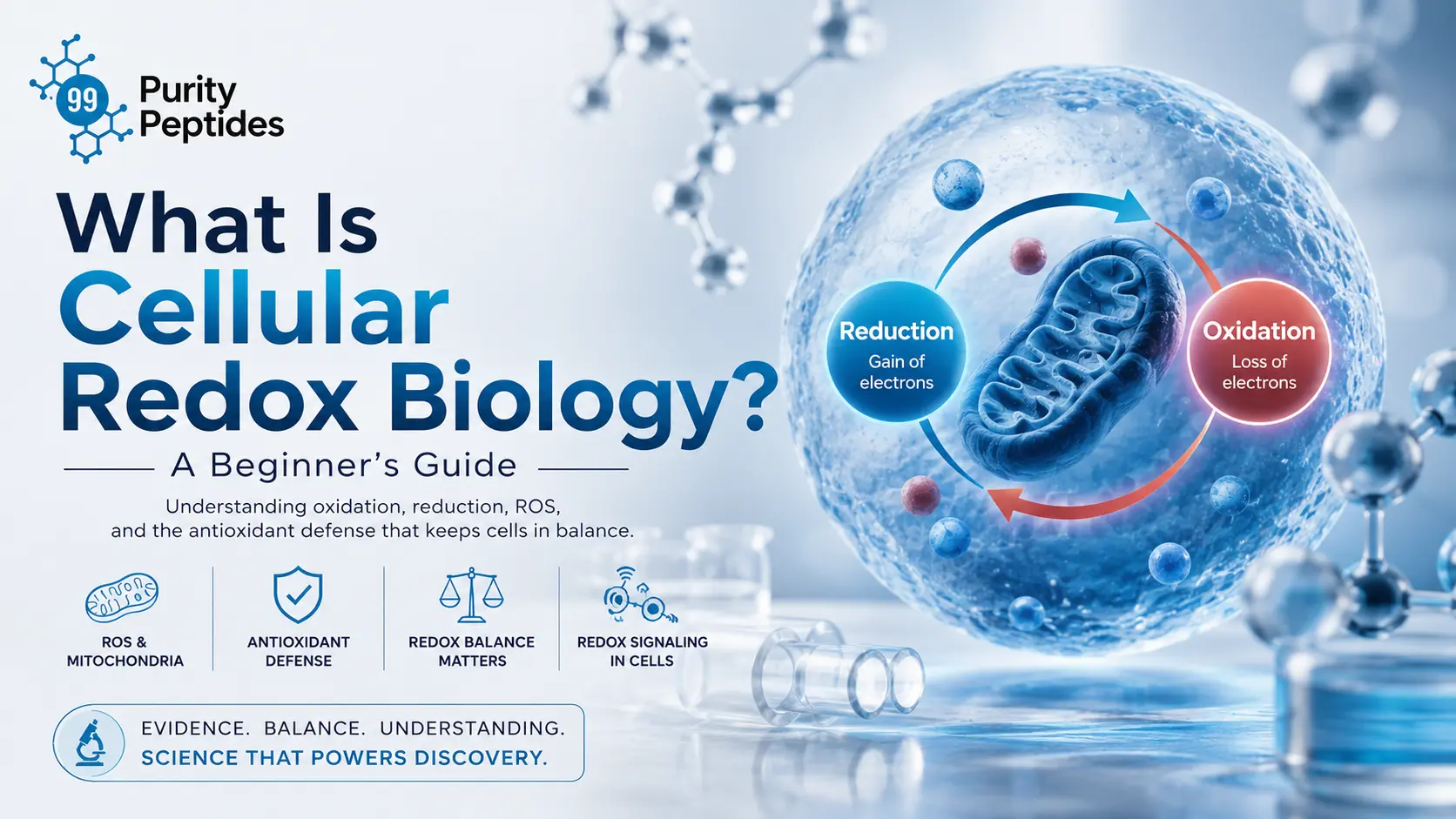
"Redox" is a portmanteau of reduction and oxidation. The two always happen together, because an electron leaving one molecule has to arrive somewhere else.
- Oxidation is the loss of electrons. The molecule that loses them is oxidized, and it acts as the reducing agent (electron donor).
- Reduction is the gain of electrons. The molecule that gains them is reduced, and it acts as the oxidizing agent (electron acceptor).
The classic mnemonic still works: OIL RIG — Oxidation Is Loss, Reduction Is Gain. The counterintuitive part is the naming. "Reduction" sounds like something is shrinking, and in a sense it is: gaining a negatively charged electron reduces the molecule’s oxidation number.
Oxidation vs. Reduction: A Side-by-Side Comparison
Feature | Oxidation | Reduction |
|---|---|---|
Electron movement | Loses electrons | Gains electrons |
Oxidation number | Increases | Decreases |
Role in the pair | Reducing agent (electron donor) | Oxidizing agent (electron acceptor) |
Common cellular example | NADH → NAD⁺ at Complex I | O₂ → H₂O at Complex IV |
Effect on the molecule | Often more reactive, less stable | Often more stable, energy-rich |
Beginner cue | "Something was taken away" | "Something was added" |
Electron Donors and Acceptors in Cells
Cells do not let electrons wander. They are handed off along defined carriers — NADH, NADPH, FADH₂, cytochrome c, coenzyme Q10 — each with a characteristic affinity for electrons. NADH and FADH₂ are the workhorse donors from cellular respiration. NADPH is kept separate on purpose: it is the reducing currency reserved almost exclusively for biosynthesis and antioxidant defense, which is why a cell can be energy-rich and still defensively bankrupt if NADPH runs low.
Redox Couples and Redox Potential
A redox couple is the oxidized and reduced form of the same molecule considered as a pair — NAD⁺/NADH, GSH/GSSG, thioredoxinₑₓ/thioredoxinᵣₑₔ. Each couple has a standard reduction potential, measured in millivolts, that describes how strongly it pulls electrons. Electrons flow spontaneously from couples with more negative potential to couples with more positive potential, which is exactly why the electron transport chain is ordered the way it is.
The redox potential of cells is not one number. Different compartments are held at deliberately different potentials — the cytosol is kept strongly reducing, the endoplasmic reticulum is comparatively oxidizing so that disulfide bonds can form in new proteins. Treating "cellular redox state" as a single global value is one of the most common beginner errors.
Reactive Oxygen Species (ROS) in Cells: What They Are and Where They Come From
Quick Answer Reactive oxygen species are oxygen-derived molecules that are chemically unstable and react quickly with lipids, proteins, and DNA. The main ones are the superoxide anion, hydrogen peroxide, and the hydroxyl radical. Most are produced as by-products of mitochondrial respiration, though enzymes also make them on purpose for signaling and immune defense. |
The Main Reactive Oxygen Species
Species | Where It Forms | Why It Matters |
|---|---|---|
Superoxide anion (O₂⁻•) | Electron leak at Complexes I and III; NADPH oxidases | The upstream precursor for most other ROS; charged, so it does not cross membranes freely |
Hydrogen peroxide (H₂O₂) | Dismutation of superoxide by SOD; peroxisomes | Relatively stable and membrane-permeable — the cell’s main redox signaling molecule |
Hydroxyl radical (•OH) | Fenton chemistry with free iron or copper | The most destructive ROS; no enzyme neutralizes it, so cells prevent it instead |
Singlet oxygen (¹O₂) | Photosensitization; some enzymatic reactions | Attacks lipids and aromatic amino acids; central to lipid peroxidation research |
Peroxynitrite (ONOO⁻) | Superoxide reacting with nitric oxide | A reactive nitrogen species; drives protein nitration and mitochondrial damage |
ROS vs. Free Radicals: What’s the Difference?
The terms are used loosely, but they are not synonyms. A free radical is any molecule with an unpaired electron — that includes superoxide and the hydroxyl radical, and also non-oxygen radicals like nitric oxide. Reactive oxygen species is a broader category defined by chemistry rather than electron pairing: hydrogen peroxide is a ROS but is not a radical, because all its electrons are paired. In short: some ROS are radicals, some radicals are not ROS, and hydrogen peroxide is the reason the distinction is worth learning.
How Reactive Oxygen Species Damage Cells
ROS do not damage cells in one way. They have three distinct target classes, and each produces a different research biomarker:
- DNA damage. Hydroxyl radicals attack guanine to form 8-oxo-dG, one of the most widely measured oxidative lesions. Unrepaired, these become mutations.
- Lipid peroxidation. Radicals abstract hydrogen from polyunsaturated fatty acids in membranes, starting a self-propagating radical chain reaction. Malondialdehyde and 4-HNE are the downstream markers.
- Protein oxidation. Cysteine and methionine residues oxidize first. Some of this is reversible and used for signaling; heavy carbonylation is not, and the protein is flagged for degradation.
Mitochondrial Redox Biology and the Electron Transport Chain
Mitochondria are called the powerhouse of the cell so often that the phrase has stopped carrying information. Here is what it actually means in redox terms. The electron transport chain is a series of four protein complexes embedded in the inner mitochondrial membrane. NADH and FADH₂ deliver electrons at Complexes I and II. Those electrons hop down the chain through coenzyme Q and cytochrome c, each step slightly more electron-hungry than the last, releasing energy that pumps protons across the membrane. ATP synthase then lets the protons back in, and the flow drives ATP synthesis. This is oxidative phosphorylation.
Why do mitochondria produce free radicals? Electron transport is efficient but not perfect. At Complexes I and III, roughly a small percentage of electrons escape the chain early and react directly with molecular oxygen, forming the superoxide anion instead of continuing to Complex IV. Superoxide leakage rises when the chain backs up — for example when there is plenty of fuel but little demand for ATP. Mitochondrial ROS production is therefore a structural feature of aerobic metabolism, not a malfunction. |
This is why mitochondrial dysfunction and oxidative stress are so tightly linked in the research literature. Damaged mitochondria leak more electrons, which produces more ROS, which damages mitochondrial DNA and membrane lipids, which impairs the chain further. That self-reinforcing loop is the core mechanism behind a large fraction of aging research and neurodegeneration research models.
Redox Homeostasis: How Cells Maintain Redox Balance
Quick Answer Redox homeostasis is the cell’s ability to hold its redox couples — mainly GSH/GSSG and NAD⁺/NADH — within a narrow operating range despite continuous ROS production. It is an active, energy-consuming steady state, not a passive equilibrium, and each cellular compartment maintains its own set point. |
Redox balance is best understood as a budget rather than a scale. On one side, ROS are generated continuously by respiration, NADPH oxidases, peroxisomal oxidases, and detoxification enzymes. On the other, a layered defense system removes them and repairs what got through. Redox imbalance occurs when production outpaces removal, or when repair capacity fails.
The NAD⁺/NADH Ratio and Redox State
The NAD⁺ to NADH ratio is one of the most informative single readouts of a cell’s redox state. A high NAD⁺/NADH ratio indicates an oxidized cytosol with capacity to accept more electrons — typically a cell actively burning fuel. A collapsed ratio signals that electron acceptors are saturated, glycolysis will stall, and superoxide leakage tends to rise. Because NAD⁺ is also the substrate for sirtuins and PARPs, this ratio links redox state directly to gene expression regulation and DNA repair, which is why it appears so often in aging research.
Antioxidant Defense Systems: How Cells Neutralize Free Radicals
Cells do not rely on dietary antioxidants for their primary defense. The heavy lifting is done by endogenous antioxidant enzymes that work at rates approaching the physical limit of diffusion — orders of magnitude faster than any vitamin.
The Main Antioxidant Enzymes
Enzyme | What It Does | Where It Works |
|---|---|---|
Superoxide dismutase (SOD) | Converts superoxide into hydrogen peroxide and oxygen | SOD1 cytosol, SOD2 mitochondrial matrix, SOD3 extracellular |
Catalase | Breaks hydrogen peroxide into water and oxygen | Mainly peroxisomes; handles high peroxide loads |
Glutathione peroxidase | Reduces hydrogen peroxide and lipid peroxides using GSH | Cytosol and mitochondria; handles low-level peroxide |
Glutathione reductase | Regenerates GSH from GSSG using NADPH | Cytosol and mitochondria; keeps the GSH pool charged |
Peroxiredoxin / Thioredoxin | Reduces peroxides via cysteine thiols; relays redox signals | All compartments; central to redox signaling |
How Antioxidant Enzymes Neutralize Free Radicals: Step by Step
- Superoxide forms when an electron escapes the electron transport chain and reacts with oxygen.
- Superoxide dismutase converts two superoxide molecules into one hydrogen peroxide and one oxygen — a controlled hand-off, not elimination.
- Glutathione peroxidase or a peroxiredoxin reduces the hydrogen peroxide to water, oxidizing two GSH molecules into one GSSG in the process.
- Glutathione reductase regenerates two GSH from GSSG, spending one NADPH.
- The pentose phosphate pathway resupplies NADPH, closing the loop and returning the cell to its baseline redox potential.
Notice the dependency chain. Antioxidant defense ultimately runs on NADPH, which runs on glucose metabolism. This is why researchers studying oxidative stress often manipulate the pentose phosphate pathway rather than adding antioxidants directly — it targets the constraint instead of the symptom.
Glutathione: The Master Antioxidant
Glutathione (GSH) is a tripeptide of glutamate, cysteine, and glycine, present in the cytosol at millimolar concentrations — extraordinarily high for a small molecule. It earns the "master antioxidant" label for three reasons: it is the substrate for glutathione peroxidase, it directly scavenges radicals, and the GSH/GSSG ratio serves as the cell’s principal redox buffer and readout. A healthy cell keeps that ratio steeply in favor of reduced GSH; a shift toward GSSG is a hallmark of oxidative stress in cell culture studies. Glutathione depletion is also one of the most reproducible findings in aging research models.
Endogenous vs. Exogenous Antioxidants
Endogenous antioxidants are made by the cell — glutathione, thioredoxin, SOD, catalase, coenzyme Q10, alpha lipoic acid. Exogenous antioxidants come from outside — vitamin C, vitamin E, dietary polyphenols. The research picture is that exogenous antioxidants mostly act as regenerators and chain-breakers rather than primary defenders. Vitamin E stops lipid peroxidation chain reactions in membranes; vitamin C then regenerates the oxidized vitamin E. Useful, but supporting cast.
The Nrf2 / Keap1 Pathway in Simple Terms
Cells do not just run a fixed defense — they scale it. Nrf2 is a transcription factor that switches on a large battery of antioxidant genes. Under normal conditions, Keap1 binds Nrf2 and tags it for continuous destruction, so almost none of it reaches the nucleus. Keap1 carries reactive cysteine residues that act as chemical sensors. When oxidants modify those cysteines, Keap1 releases its grip, Nrf2 accumulates, moves to the nucleus, and binds the antioxidant response element in target gene promoters. The result is more SOD, more glutathione synthesis, more detoxification enzymes. Keap1 is the sensor, Nrf2 is the switch, and the antioxidant response element is the destination.
Redox Signaling Pathways: When Oxidation Is a Message, Not Damage
This is the section that separates modern redox biology from the 1990s "free radicals are bad" framing. Cells deliberately generate ROS to carry information. Growth factor receptors trigger localized hydrogen peroxide production. That peroxide reversibly oxidizes a cysteine on a nearby phosphatase, temporarily disabling it and allowing a phosphorylation signal to proceed. The oxidation is specific, local, brief, and reversible — the defining features of a signal rather than damage. Redox-sensitive transcription factors including NF-κB and the MAPK and PI3K/Akt pathways all respond to these cues, which is why redox signaling and inflammation are so entangled in the literature.
Oxidative Eustress vs. Oxidative Distress
Dimension | Oxidative Eustress | Oxidative Distress |
|---|---|---|
ROS level | Low, physiological | High, sustained |
Location | Localized, compartment-specific | Widespread across the cell |
Duration | Brief and self-limiting | Chronic |
Molecular effect | Reversible cysteine oxidation | Irreversible oxidation, strand breaks, carbonylation |
Cellular outcome | Adaptation, signaling, hormesis | Damage, senescence, cell death |
Research relevance | Exercise adaptation, Nrf2 activation studies | Disease models, aging biomarkers |
Hormesis is the principle underneath this table: a mild stressor triggers an adaptive response that leaves the cell more resilient than before. It also explains one of the most cited surprises in the field — high-dose antioxidant intervention studies have repeatedly failed to produce the benefits the free radical theory predicted. Blanket suppression of ROS also suppresses the signals that trigger adaptation. Is oxidative stress always harmful? The evidence says no, and that answer reshaped the discipline.
Oxidative Stress, Aging, and Disease Research
The Free Radical Theory of Aging, Explained Simply
Proposed by Denham Harman in 1956, the free radical theory of aging holds that aging results from cumulative oxidative damage to cellular components, primarily from mitochondrial ROS. It elegantly explained why metabolic rate, mitochondria, and lifespan seem related across species.
The theory has since been substantially revised rather than abandoned. Several findings complicated it: some long-lived species produce ROS at high rates, some antioxidant-overexpressing model organisms show no lifespan extension, and mild mitochondrial stress can extend lifespan in several models. The contemporary view treats redox imbalance as one interacting hallmark of aging alongside cellular senescence, telomere attrition, and proteostasis failure — significant, but not the single master cause.
Where Redox Biology Shows Up in Disease Research
- Neurodegeneration research — neurons combine high oxygen consumption, high lipid content, and limited replacement capacity, a difficult combination for redox management.
- Cardiovascular research — ischemia–reperfusion injury is a textbook oxidative burst, and lipid peroxidation is central to atherosclerosis models.
- Cancer cell metabolism — tumor cells often run at elevated basal ROS that drives proliferative signaling while depending heavily on antioxidant capacity to survive it, which makes redox state a studied vulnerability.
- Metabolic disease research — mitochondrial dysfunction and redox imbalance appear consistently in insulin resistance models.
- Inflammation research — NADPH oxidase-derived ROS are both weapons and signals in immune activation.
How Researchers Study Redox Biology in the Lab
How Redox Potential Is Measured
- Select the readout. Decide whether you are measuring a redox couple ratio (GSH/GSSG, NAD⁺/NADH), a specific ROS, or downstream damage.
- Choose the probe. Genetically encoded sensors such as roGFP and HyPer report compartment-specific redox state in living cells; small-molecule fluorescent probes are simpler but less specific.
- Control the sampling. Redox couples re-equilibrate within seconds of lysis, so samples are typically quenched immediately and thiols alkylated to prevent artifactual oxidation.
- Quantify. Spectrophotometry, HPLC, mass spectrometry, or plate-reader fluorescence, depending on the analyte and required sensitivity.
- Validate with an orthogonal method. Fluorescent ROS probes are notoriously prone to artifacts; a second, chemically unrelated method is expected in credible work.
Common Biomarkers of Oxidative Stress
- 8-oxo-dG — oxidative DNA damage
- Malondialdehyde (MDA) and 4-HNE — lipid peroxidation
- Protein carbonyls — irreversible protein oxidation
- GSH/GSSG ratio — overall redox buffer status
- F2-isoprostanes — widely regarded as among the more reliable lipid peroxidation markers
- Antioxidant enzyme activity assays — SOD, catalase, glutathione peroxidase
Common Techniques
Cell culture assays for controlled ROS exposure, Western blot for oxidized or Nrf2-target proteins, ELISA for biomarker quantification, spectrophotometry for enzyme kinetics, and HPLC or mass spectrometry for precise measurement of glutathione and nucleotide pools. Most redox work combines at least three of these, because no single method is trusted alone.
How Peptides Are Used in Redox and Mitochondrial Research
Peptides show up throughout redox biology research for a structural reason: many of the cell’s own redox machinery components are peptides and proteins built around reactive cysteine thiols. Glutathione itself is a tripeptide. Synthetic peptides give researchers a way to probe cellular repair mechanisms, mitochondrial targeting, and cell signaling pathways with defined amino acid sequences and known molecular weight, rather than the mixed composition of a natural extract.
In practice, research peptides appear in redox and mitochondrial research as tools for in vitro studies of signaling pathway activation, mitochondrial membrane interaction, antioxidant enzyme expression, and cell viability under induced oxidative stress. The value is experimental control: a defined sequence, a verified purity, and a reproducible input.
Research Use Only All peptides referenced by 99 Purity Peptides are supplied strictly as laboratory research compounds. They are not for human consumption, not for diagnostic use, and not for therapeutic use. No dosing, administration, or self-use guidance is provided anywhere on this site. Handling and disposal are the responsibility of the qualified researcher. |
What Is Considered High Purity for Research Peptides?
In practice, research-grade peptides are typically specified at 98%+ purity as measured by HPLC, with mass spectrometry confirming the molecular weight matches the intended amino acid sequence. Purity alone is not the whole picture — peptide solubility, peptide stability, and storage stability all affect whether a lyophilized peptide vial actually performs as specified months after it arrives.
How to Choose a Reliable Research Peptide Supplier
- Insist on a certificate of analysis for the specific lot you receive — not a generic sample document for the product line.
- Confirm both HPLC purity analysis and mass spectrometry identity confirmation appear on that COA.
- Check for independent third-party testing rather than in-house numbers alone.
- Verify batch-to-batch consistency, since reproducibility across experiments depends on it.
- Review storage and shipping conditions — lyophilized peptides shipped without appropriate handling can degrade before first use.
- Confirm the supplier labels products clearly for research use only. Vague or health-implying language is a compliance warning sign, not a marketing style.
Research-Grade vs. Pharmaceutical-Grade Peptides
Dimension | Research-Grade | Pharmaceutical-Grade |
|---|---|---|
Intended use | Laboratory and in vitro research only | Clinical use in humans |
Regulatory oversight | Labeled as a research chemical; not FDA-approved for human use | Manufactured under GMP and subject to regulatory approval |
Typical documentation | Certificate of analysis, HPLC and MS data | Full GMP batch records and regulatory filings |
Sterility requirements | Not required to be sterile or pyrogen-free | Sterility and endotoxin limits enforced |
Appropriate context | Cell culture studies, biomarker analysis, method development | Patient treatment under medical supervision |
Common Beginner Mistakes in Redox Biology
- Treating all ROS as identical. Hydrogen peroxide is a signal; the hydroxyl radical is pure damage. Conflating them makes most papers unreadable.
- Assuming more antioxidants is always better. Suppressing redox signaling can block the adaptive responses you were trying to study.
- Reporting a single "cellular redox state." Compartments differ by design; specify which one.
- Trusting one fluorescent probe. Probe artifacts are among the best-documented reproducibility problems in the field.
- Confusing correlation with mechanism. Elevated oxidative stress biomarkers accompany many disease models without proving causation.
- Ignoring reagent provenance. Undocumented compound purity is one of the quietest sources of irreproducible redox data.
Key Takeaways
- Cellular redox biology studies how cells transfer electrons through paired oxidation and reduction reactions to make energy, signal, and defend themselves.
- Oxidation is electron loss; reduction is electron gain. They always occur together as a redox couple.
- Mitochondria produce reactive oxygen species as an unavoidable by-product of the electron transport chain, not as a malfunction.
- Hydrogen peroxide is the cell’s main redox signaling molecule; the hydroxyl radical is the most destructive ROS and has no dedicated enzyme.
- Redox homeostasis is an active, compartment-specific steady state, tracked most usefully through the GSH/GSSG and NAD⁺/NADH ratios.
- Antioxidant enzymes — SOD, catalase, glutathione peroxidase, peroxiredoxins — carry the primary defense, and ultimately depend on NADPH.
- The Nrf2/Keap1 pathway lets cells scale their antioxidant defenses in response to oxidant signals.
- Oxidative eustress and oxidative distress are chemically similar but functionally opposite — dose, duration, and location decide which one you get.
- The free radical theory of aging has been revised, not discarded; redox imbalance is one interacting hallmark among several.
- In research settings, compound quality matters as much as experimental design — verified purity, a lot-specific COA, and third-party testing are baseline requirements.
Frequently Asked Questions
What is cellular redox biology in simple terms?
Cellular redox biology is the study of how cells transfer electrons between molecules through paired oxidation and reduction reactions. Those transfers power energy production, carry signals between proteins, and drive the antioxidant defenses that keep cells stable.
What does "redox" actually mean?
Redox is short for reduction-oxidation. It describes any reaction where one molecule loses electrons (oxidation) and another gains them (reduction). The two halves always occur together, because electrons cannot simply disappear.
Why is redox balance important for healthy cells?
Redox balance determines whether oxidants act as signals or as damage. Within its normal range, controlled oxidation regulates gene expression and adaptation. Outside it, the same molecules damage DNA, membrane lipids, and proteins.
What are reactive oxygen species (ROS)?
Reactive oxygen species are unstable oxygen-derived molecules including the superoxide anion, hydrogen peroxide, and the hydroxyl radical. Most form as by-products of mitochondrial respiration, though some enzymes produce them deliberately for signaling and immune defense.
What causes oxidative stress in cells?
Oxidative stress occurs when ROS production outpaces antioxidant capacity. Common research-model causes include mitochondrial dysfunction, chronic inflammation, depleted glutathione, free iron driving Fenton chemistry, radiation, and toxin exposure.
What is the difference between ROS and free radicals?
A free radical has an unpaired electron; ROS is a chemistry-based category of oxygen-derived reactive molecules. Hydrogen peroxide is a ROS but not a radical, while nitric oxide is a radical but not a ROS.
What role do mitochondria play in redox biology?
Mitochondria run the electron transport chain, which generates most cellular ATP and also leaks a fraction of its electrons to oxygen, forming superoxide. They are simultaneously the cell’s largest energy source and its largest ROS source.
What is glutathione and why is it important?
Glutathione (GSH) is a tripeptide antioxidant present at millimolar concentrations in the cytosol. It supplies electrons to glutathione peroxidase, scavenges radicals directly, and its GSH/GSSG ratio serves as the cell’s main redox buffer and readout.
What is the Nrf2 antioxidant pathway?
Nrf2 is a transcription factor normally held inactive by Keap1. When oxidants modify Keap1’s reactive cysteines, Nrf2 is released, enters the nucleus, and activates antioxidant response element genes, increasing the cell’s antioxidant enzyme production.
What is oxidative eustress versus oxidative distress?
Oxidative eustress is low-level, localized, reversible oxidation used for signaling and adaptation. Oxidative distress is high, sustained, widespread oxidation that causes irreversible damage. The chemistry is similar; dose, duration, and location differ.
How is redox status measured in research studies?
Researchers measure redox couple ratios such as GSH/GSSG and NAD⁺/NADH, use genetically encoded probes like roGFP and HyPer for live-cell readouts, and quantify damage biomarkers by HPLC, mass spectrometry, ELISA, or spectrophotometry.
What is the free radical theory of aging?
Proposed by Denham Harman in 1956, it holds that aging results from accumulated oxidative damage, largely from mitochondrial ROS. It has since been revised: redox imbalance is now treated as one interacting hallmark of aging rather than the single cause.
What is the NAD⁺/NADH ratio and why does it matter?
It compares oxidized to reduced nicotinamide adenine dinucleotide and indicates a cell’s capacity to accept more electrons. A high ratio reflects active oxidative metabolism; a collapsed ratio signals stalled electron flow and rising ROS leakage.
How do peptides relate to redox and mitochondrial research?
Synthetic peptides give researchers defined-sequence tools for in vitro studies of redox signaling, mitochondrial interaction, and antioxidant enzyme expression. Their value is experimental control — known composition, verified purity, reproducible results.
What should I look for in a research peptide supplier?
Look for a lot-specific certificate of analysis, HPLC purity data, mass spectrometry identity confirmation, independent third-party testing, documented storage and shipping conditions, and clear research-use-only labeling on every product.
Are 99 Purity Peptides products intended for human consumption?
No. All products are supplied strictly for laboratory research use. They are not intended for human or veterinary consumption, diagnostic use, or therapeutic use, and no dosing or administration guidance is provided.